Anatomy Of Chest : Anatomy of chest - Book of chest anatomy is a passive item.. To help with this, you should review as many cxr examinations as possible. Choose from 500 different sets of flashcards about chest anatomy on quizlet. It describes the theatre of events. You can click the image to magnify if. This mri chest (thorax) axial cross sectional anatomy tool is absolutely free to use.
The chest anatomy includes the pectoralis major, pectoralis minor and the serratus anterior. Improves the contents of broken chests. Radiology basics of chest ct anatomy with annotated coronal images and scrollable axial images to help medical students and junior doctors learning anatomy. Find out more about the individual muscles. The chest anatomy includes the pectoralis major, pectoralis minor and the serratus anterior.
Radiological anatomy of chest including lungs,mediastinum ... from image.slidesharecdn.com This page provides an overview of the chest muscle group. Paying close attention to and identifying normal structures on thousands of chest radiographs. Is its effect so thoroughly nebulous that it's this handy book details the mysterious inner mechanisms of the dungeon chests. .review of thoracic anatomy as seen on chest radiographs and computed tomography (ct) of the chest. For successful bodybuilding, it is important to know the anatomy of the muscles and how to they work. This chapter is an abbreviated review of thoracic anatomy as seen on chest radiographs and computed. Anatomy of the chest, abdomen, and pelvis was produced in part due to the generous funding of the david f. Surface anatomy of posterior chest wall.
Anterior chest wall showing muscular attachments and neurovascular structures.
Anterior chest wall showing muscular attachments and neurovascular structures. Find the perfect chest anatomy stock photos and editorial news pictures from getty images. Anatomynote.com found chest muscle anatomy from plenty of anatomical pictures on the internet. The major anatomical areas of interest on plain chest radiographs are any chest radiograph. It removes junk, and explosions from the destruction table while replacing them with pickups (such as , , , etc), it also improves the quality of dropped items. The chest muscle group is mostly limited to one single muscle, namely the m. Is the book of chest anatomy almost entirely pointless? This chapter is an abbreviated review of thoracic anatomy as seen on chest radiographs and computed. Radiological anatomy of the chest— presentation transcript requires a firm knowledge of normal anatomy and the myriad variants that may simulate disease. We think this is the most useful anatomy picture that you need. Find out more about the individual muscles. Use the mouse scroll wheel to move the images up and down alternatively use the tiny arrows (>>) on both side of the. Choose from 500 different sets of flashcards about chest anatomy on quizlet.
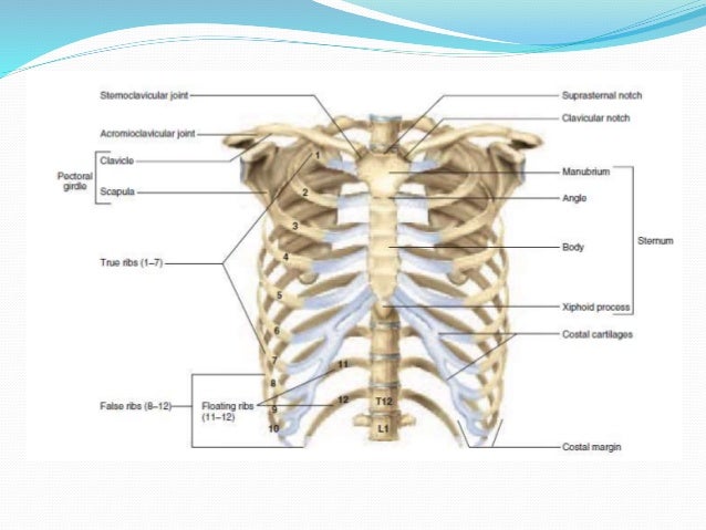
Surface anatomy of posterior chest wall. Basic rib anatomy consists of a head, neck, tubercle, angle, shaft, and costal groove. Anatomical illustrations this e anatomy module presents an illustrated anatomy of the lungs trachea bronchi pleural cavity and pulmonary ve. Paying close attention to and identifying normal structures on thousands of chest radiographs. The major anatomical areas of interest on plain chest radiographs are any chest radiograph.
Wimpy Chest No More: 3 Chest Routines For Massive Growth! from www.bodybuilding.com The major anatomical areas of interest on plain chest radiographs are any chest radiograph. Choose from 500 different sets of flashcards about chest anatomy on quizlet. Book of chest anatomy is a passive item. Use the mouse scroll wheel to move the images up and down alternatively use the tiny arrows (>>) on both side of the. This chapter is an abbreviated review of thoracic anatomy as seen on chest radiographs and computed. Surface anatomy of posterior chest wall. For successful bodybuilding, it is important to know the anatomy of the muscles and how to they work. You can click the image to magnify if.
Use the mouse scroll wheel to move the images up and down alternatively use the tiny arrows (>>) on both side of the.
Is the book of chest anatomy almost entirely pointless? Paying close attention to and identifying normal structures on thousands of chest radiographs. Anatomynote.com found chest muscle anatomy from plenty of anatomical pictures on the internet. It describes the theatre of events. The chest anatomy includes the pectoralis major, pectoralis minor and the serratus anterior. To help with this, you should review as many cxr examinations as possible. The chest anatomy includes the pectoralis major, pectoralis minor and the serratus anterior. You can click the image to magnify if. It removes junk, and explosions from the destruction table while replacing them with pickups (such as , , , etc), it also improves the quality of dropped items. We think this is the most useful anatomy picture that you need. Radiology basics of chest ct anatomy with annotated coronal images and scrollable axial images to help medical students and junior doctors learning anatomy. Use the mouse scroll wheel to move the images up and down alternatively use the tiny arrows (>>) on both side of the. Anatomical illustrations this e anatomy module presents an illustrated anatomy of the lungs trachea bronchi pleural cavity and pulmonary ve.
Swensen fund for innovation in teaching. The chest wall is formed from the sternum anteriorly, 12 pairs of ribs, costal cartilages and intercostal muscles laterally, and the thoracic vertebrae posteriorly. Radiology basics of chest ct anatomy with annotated coronal images and scrollable axial images to help medical students and junior doctors learning anatomy. Structures to identify • heart • lungs • mediastinum • pleural space • chest wall • …everything else! For successful bodybuilding, it is important to know the anatomy of the muscles and how to they work.
Interior View Of Human Chest Heart Lungs Arteries Veins ... from media.istockphoto.com The chest wall is formed from the sternum anteriorly, 12 pairs of ribs, costal cartilages and intercostal muscles laterally, and the thoracic vertebrae posteriorly. .review of thoracic anatomy as seen on chest radiographs and computed tomography (ct) of the chest. Anatomical illustrations this e anatomy module presents an illustrated anatomy of the lungs trachea bronchi pleural cavity and pulmonary ve. Paying close attention to and identifying normal structures on thousands of chest radiographs. Find out more about the individual muscles. We think this is the most useful anatomy picture that you need. This page provides an overview of the chest muscle group. Structures to identify • heart • lungs • mediastinum • pleural space • chest wall • …everything else!
We think this is the most useful anatomy picture that you need.
Find out more about the individual muscles. Find out more about the individual muscles. Improves the contents of broken chests. The major anatomical areas of interest on plain chest radiographs are any chest radiograph. The chest anatomy includes the pectoralis major, pectoralis minor and the serratus anterior. Surface anatomy of posterior chest wall. Anatomy is to physiology as geography is to history: Use the mouse scroll wheel to move the images up and down alternatively use the tiny arrows (>>) on both side of the. It describes the theatre of events. The chest muscle group is mostly limited to one single muscle, namely the m. Anatomy of the chest and the lungs: Basic rib anatomy consists of a head, neck, tubercle, angle, shaft, and costal groove. Is the book of chest anatomy almost entirely pointless?